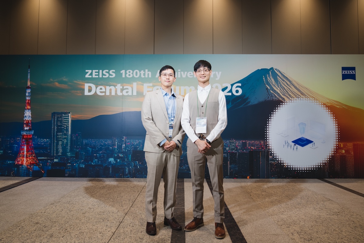
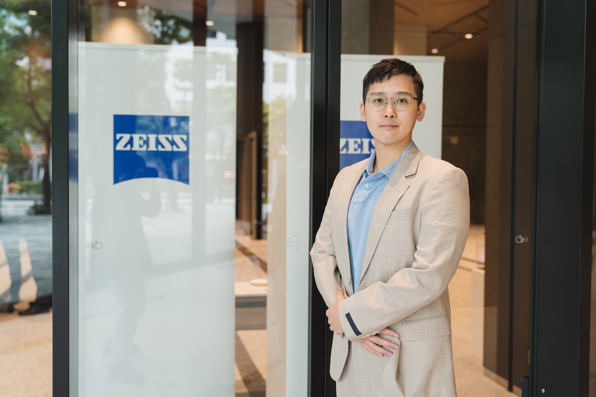
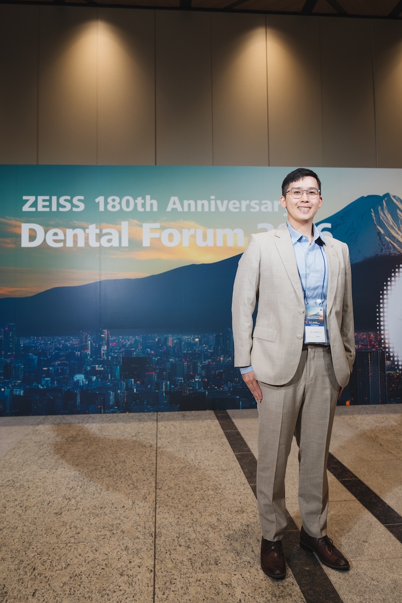
全球光學知名品牌蔡司(ZEISS)於今年正式迎來創立 180 週年的重要里程碑。身為醫療顯微技術與光學透鏡發展的代表企業,蔡司特別在日本東京舉行「ZEISS Dental Forum 2026」國際學術論壇。這場會議吸引了來自日本、亞洲及世界各地的牙科專科醫師、學者與臨床專家共同參與,針對顯微牙科的臨床實務、微創治療理念與前沿技術進行交流。
The world-renowned optical brand ZEISS officially celebrates its 180th anniversary milestone this year. As a benchmark enterprise in medical microscopy and optical lens development, ZEISS held the "ZEISS Dental Forum 2026" international academic forum in Tokyo, Japan. The event brought together dental specialists, scholars, and clinical experts from Japan, Asia, and around the globe to exchange insights on microscopic dental clinical practice, minimally invasive treatment concepts, and cutting-edge technologies.
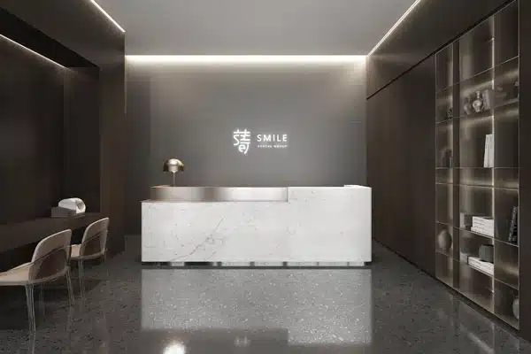
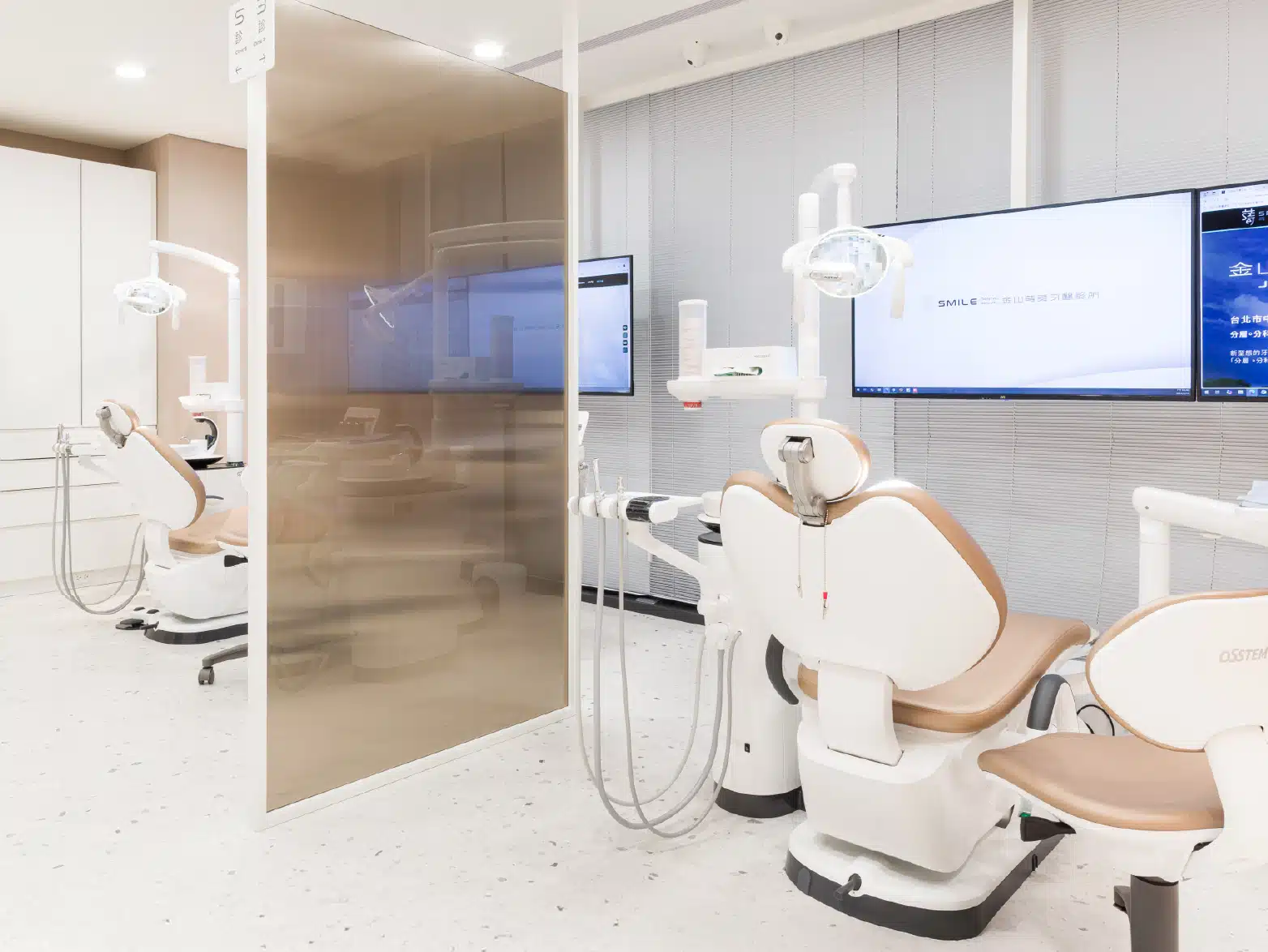
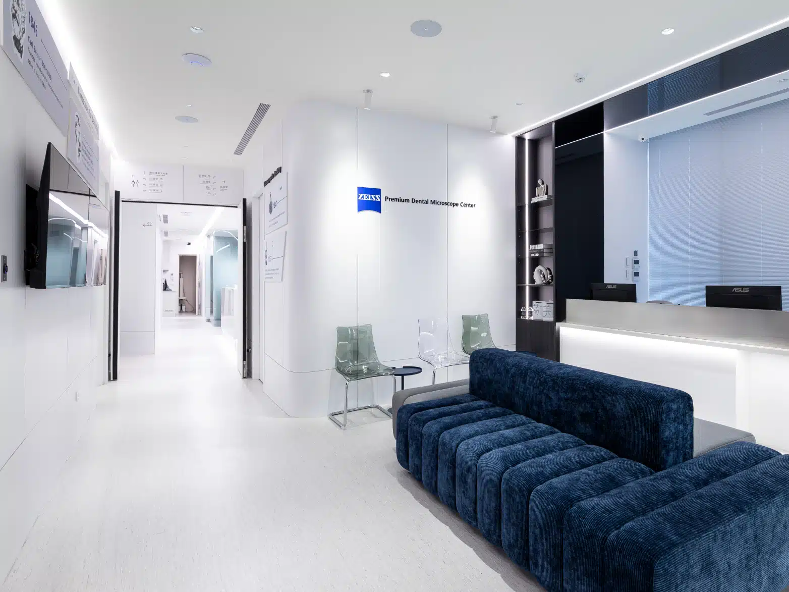
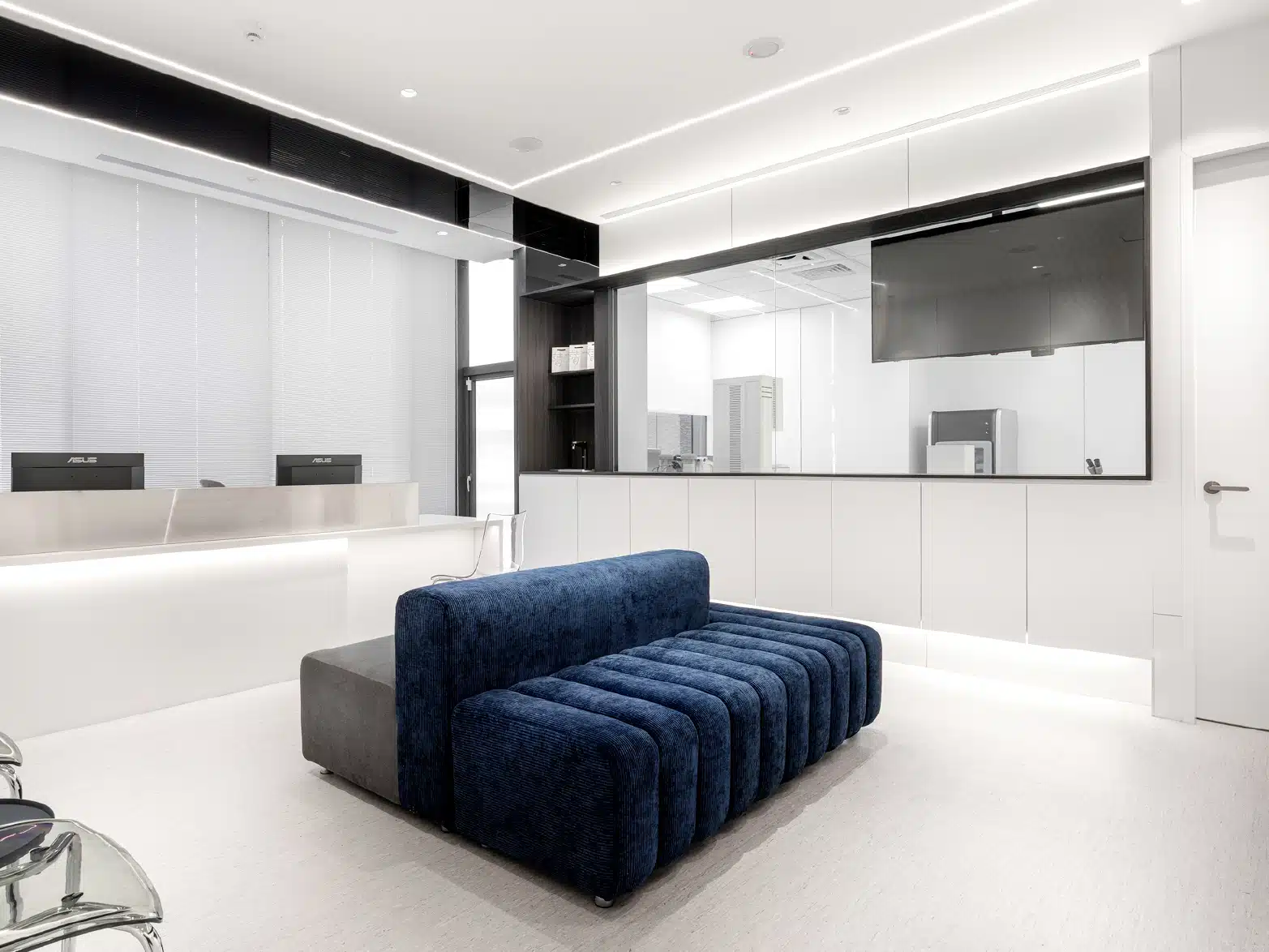
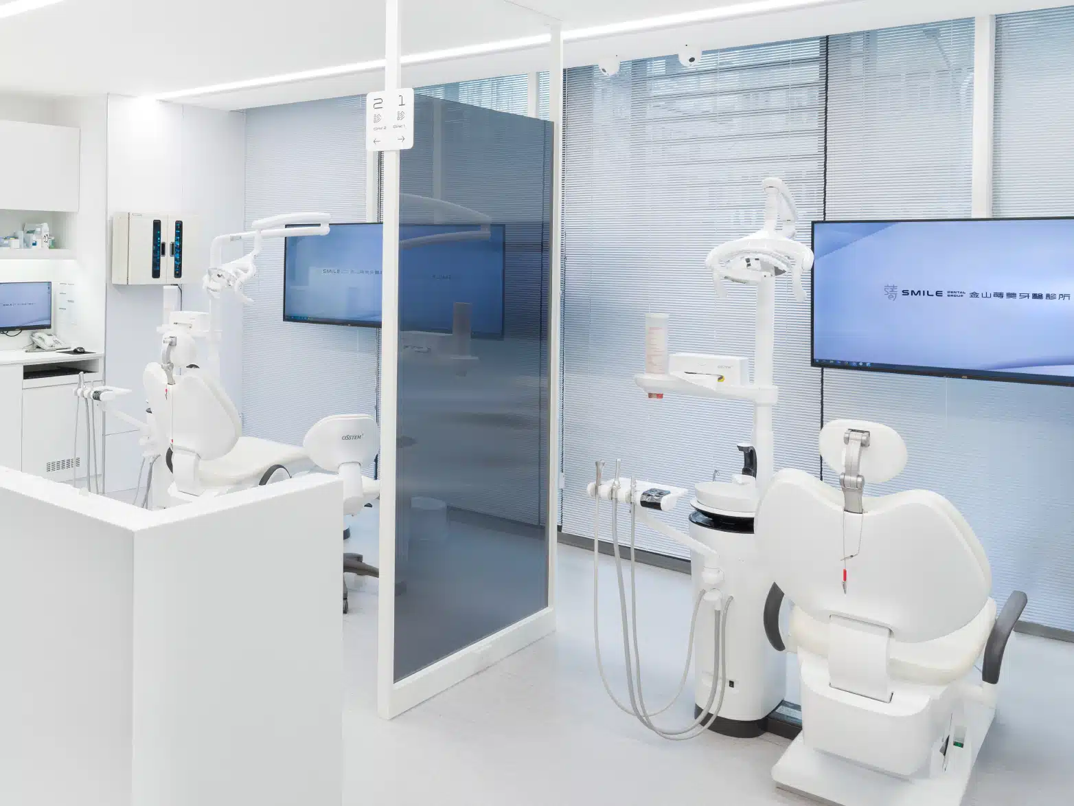
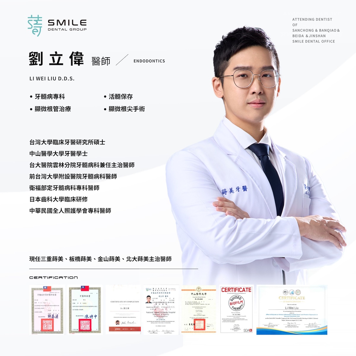
長期關注數位化與顯微專科發展的「蒔美牙醫集團」,持續透過參與國際學術會議,將各國臨床經驗與醫療觀點帶回診間。在牙齒深層的根管治療領域,蒔美牙醫集團由顯微鏡根管專科醫師劉立偉醫師主持,善用高倍率顯微鏡輔助,針對複雜的根管病灶進行細緻的評估與處置,協助患者評估保留自然牙結構的可能。
Smile Dental Group, which has long focused on the development of digital dentistry and microscopic specialties, continuously brings international clinical experiences and medical perspectives back to its clinics by participating in global academic conferences. In deep root canal treatment, Smile Dental Group is led by endodontic specialist Dr. Liu Li Wei, who utilizes high-magnification microscopes for meticulous evaluation and treatment of complex root canal lesions, helping patients assess the potential for preserving their natural tooth structure.
直擊東京 ZEISS Dental Forum 2026:高倍率視野帶動微創醫療觀點
Inside the Tokyo ZEISS Dental Forum 2026: High-Magnification Vision Driving Minimally Invasive Concepts
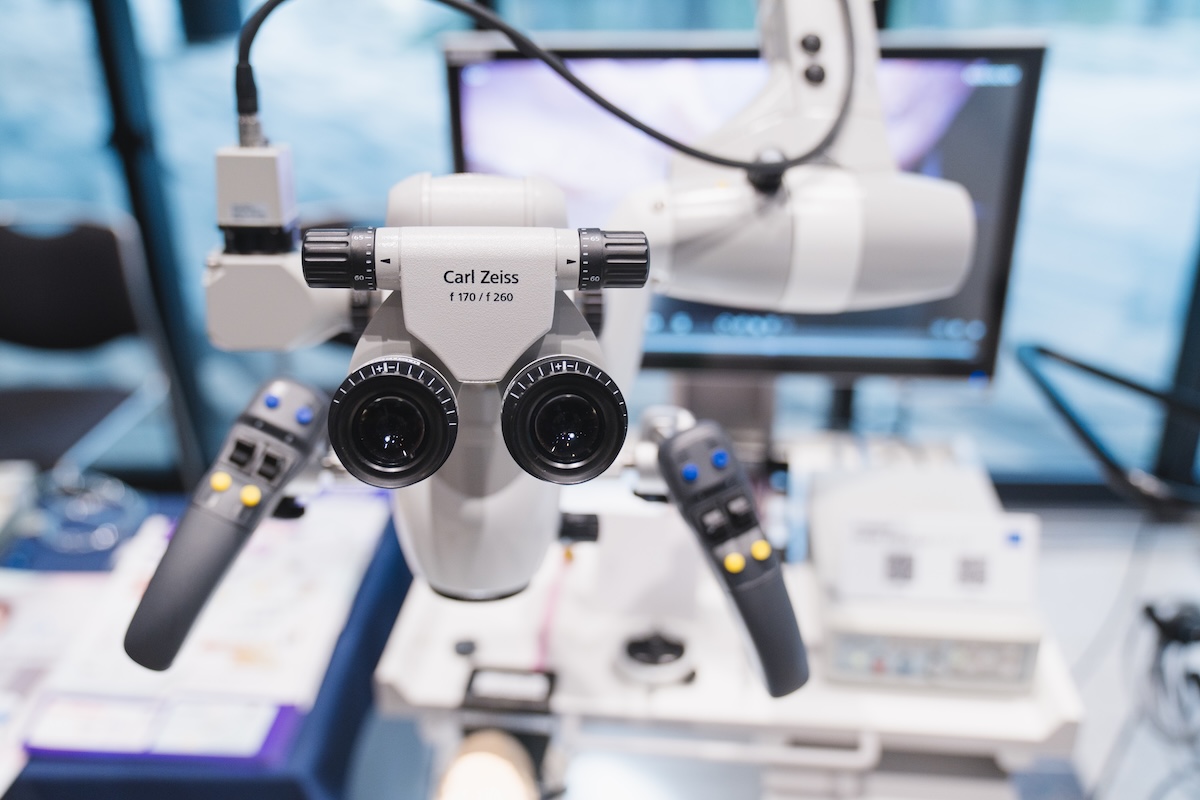
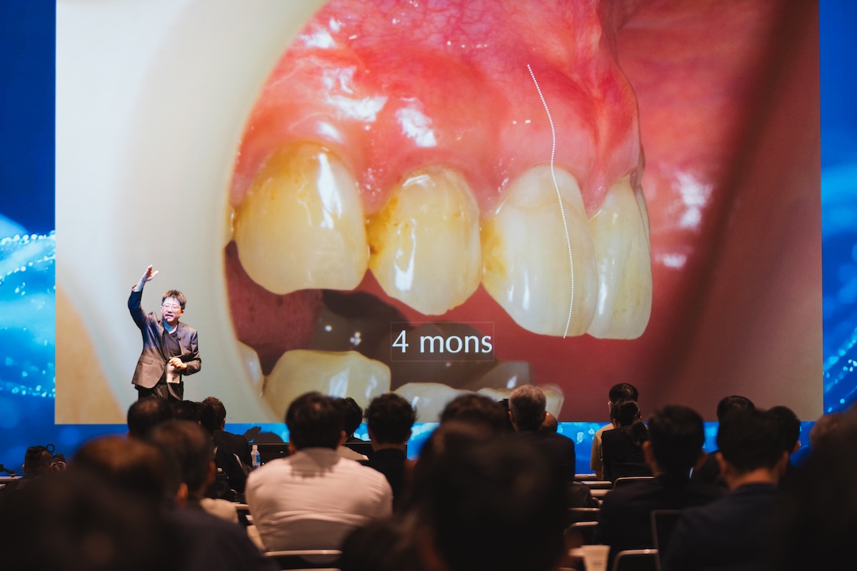
在「ZEISS Dental Forum 2026」東京論壇中,研討內容圍繞著「高倍率放大視野」、「齒質保留」與「臨床療效穩定」等核心議題。現代牙科醫療逐漸強調微創理念,透過顯微鏡的輔助,臨床醫師得以在顯微視野下檢視細微結構,改變了過往主要依賴目視與經驗的臨床模式。
At the "ZEISS Dental Forum 2026" in Tokyo, discussions centered on core topics such as "high-magnification vision," "tooth structure preservation," and "clinical stability." Modern dentistry increasingly emphasizes minimally invasive concepts. With the aid of microscopes, clinicians can inspect subtle structures under microscopic vision, shifting away from traditional clinical models that relied primarily on direct line-of-sight and experience.
牙齒內部的解剖構造極為微小且複雜,尤其是位於牙齒最深處的牙髓根管系統,其管道直徑常僅有數百微米。在傳統治療中,醫師常面臨視野受限或光線不足的問題;高倍率顯微技術的發展,則能提供清晰的照明與放大視野,協助醫師在臨床操作時擁有更明確的參考依據。
The internal anatomical structure of teeth is extremely tiny and complex, particularly the root canal system deep within the tooth, where canal diameters often measure only a few hundred micrometers. In traditional treatment, clinicians frequently face limited vision or inadequate lighting. The development of high-magnification microscopy provides bright illumination and expanded vision, giving doctors a clearer reference during clinical procedures.
蒔美牙醫集團透過參與國際學術交流,持續將相關臨床觀點與設備應用整合至診療流程中,期望在客觀、嚴謹的醫療基礎上,提供符合患者需求的照護品質。
By engaging in international academic exchanges, Smile Dental Group continuously integrates relevant clinical insights and equipment applications into its diagnostic and treatment workflows, aiming to deliver patient-centered care based on objective and rigorous medical standards.
專注牙髓病症處置:顯微鏡根管專科醫師 劉立偉醫師
Focusing on Endodontic Care: Endodontic Specialist Dr. Liu Li Wei
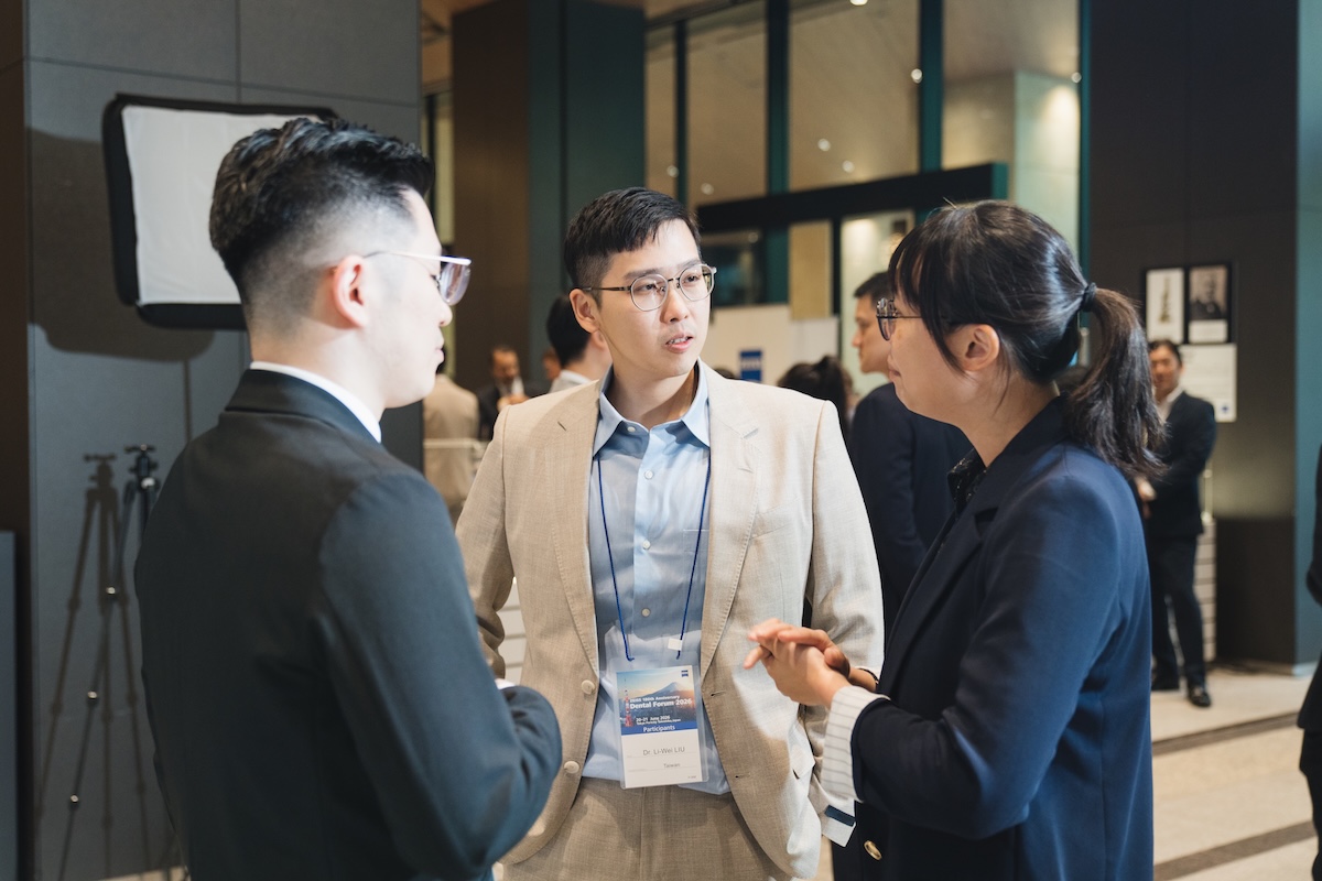
牙齒的健康與牙根結構息息相關。當牙髓組織因深度蛀牙、裂齒或外傷而受到感染或壞死時,妥善的根管治療(俗稱抽神經)是清潔根管內部並評估保留牙齒的重要處置。若根管清潔不夠完善,殘留的細菌可能持續引發根尖周圍組織發炎,甚至影響牙周健康。
Tooth health is closely tied to root structure. When dental pulp tissue becomes infected or necrotic due to deep cavities, cracked teeth, or trauma, proper root canal treatment is essential for cleaning the canal interior and evaluating tooth retention. If root canal cleaning is incomplete, residual bacteria may continue to trigger periapical tissue inflammation, potentially compromising periodontal health.
顯微鏡根管專科劉立偉醫師專注於複雜根管病灶的處置。劉立偉醫師認為,天然牙的結構與功能具有其獨特性,因此在臨床治療上,始終秉持著「盡可能保留天然齒質」的原則,藉由蔡司顯微鏡的輔助,在顯微視野下進行細致的清創與封填程序。
Endodontic specialist Dr. Liu Li Wei focuses on managing complex root canal lesions. Dr. Liu Li Wei believes that natural teeth possess unique structural and functional values. Therefore, in clinical treatment, he adheres to the principle of "preserving natural tooth structure whenever possible," performing meticulous debridement and obturation under microscopic vision with the assistance of ZEISS microscopes.
顯微視野下的臨床應用:多元複雜根管狀況的處置之道
Clinical Applications Under Microscopic Vision: Managing Diverse and Complex Root Canal Conditions
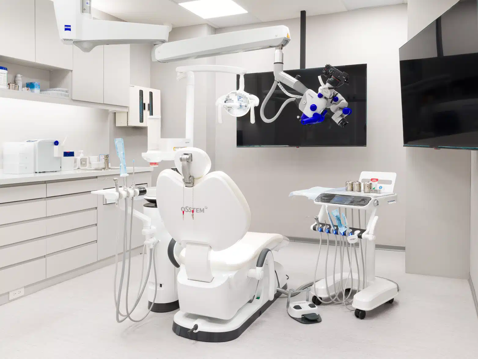
人類牙齒的根管系統並非簡單的單一管線,而是包含許多分支、彎曲甚至鈣化的複雜結構。透過蔡司顯微鏡的高倍率放大功能與同軸無影光源,原本狹窄暗沉的牙髓腔內部構造得以呈現得更加清楚,進而協助處置過往較難檢視的臨床狀況:
The human root canal system is far from a simple single pathway; it consists of complex structures with branches, curvatures, and calcifications. Through high-magnification and shadowless coaxial illumination of ZEISS microscopes, the narrow, dark internal anatomy of the pulp cavity is rendered clearly visible, helping manage clinical situations that were previously difficult to inspect:
尋找解剖變異與隱密根管
許多牙齒(例如上顎大臼齒的 MB2 根管、下顎大臼齒的 C 型根管)存在解剖結構上的變異或隱密的分支管路。若這些管路未在治療中被發現並妥善處置,感染源可能持續殘留。透過顯微放大視野,機能更明確地定位這些微小管路,進行逐步的清創與消毒。
Locating Anatomical Variations and Hidden Canals: Many teeth (such as the MB2 canal in maxillary molars or C-shaped canals in mandibular molars) feature anatomical variations or hidden branch canals. If these canals are not detected and managed during treatment, sources of infection may persist. Microscopic magnification enables doctors to locate these tiny canals more accurately for systematic debridement and disinfection.
處置根管鈣化與狹窄問題
隨著年齡增長或因長期慢性發炎,牙髓腔與根管可能出現鈣化堵塞的現象,增加治療器械進入根尖的難度。結合顯微鏡的清晰視野與超音波器械,可以在盡量少破壞健康齒質的前提下,逐步修整鈣化組織,嘗試引導管路暢通。
Managing Calcified and Narrow Canals: With aging or long-term chronic inflammation, the pulp cavity and root canals may develop calcified blockages, making instrument negotiation to the apex more challenging. Combining microscopic vision with ultrasonic instruments allows clinicians to reshape calcified tissues step-by-step while minimizing damage to healthy tooth structure, aiming to re-establish canal patency.
器械分離與牙根穿孔之評估修復
在過往的根管處置中,若遇到治療器械分離於根管內,或牙根因病變而出現穿孔,常增加處置的複雜度。經由顯微鏡的觀察,能評估以微創方式取出分離器械的可行性;對於牙根穿孔部位,則可運用生醫級材料(如 MTA 生物陶瓷)進行密封填補,提供結構復原的條件。
Evaluating and Repairing Separated Instruments and Root Perforations: In traditional root canal procedures, separated instruments within canals or root perforations caused by lesions significantly increased treatment complexity. Under microscopic inspection, doctors can evaluate the feasibility of retrieving separated instruments using minimally invasive techniques. For root perforations, biomedical materials (such as MTA bioceramics) can be applied for sealed repair, creating favorable conditions for structural recovery.
二次根管治療(根管再治療)評估
對於過去曾接受過治療但後續再次出現症狀或根尖發炎的患牙,可經由顯微鏡重新檢視牙髓腔內部,移除先前的充填物,尋找可能的發炎原因並再次進行清創,為患者提供重新評估與治療的機會。
Evaluating Retreatment (Secondary Root Canal Treatment): For teeth that received prior treatment but later exhibit symptoms or periapical inflammation, doctors can re-examine the interior pulp chamber under a microscope, remove old filling materials, identify underlying causes of inflammation, and perform re-cleansing—offering patients a chance for re-evaluation and retreatment.
顯微根管治療的臨床特點與評估效益
Clinical Features and Evaluation Benefits of Microscopic Endodontics
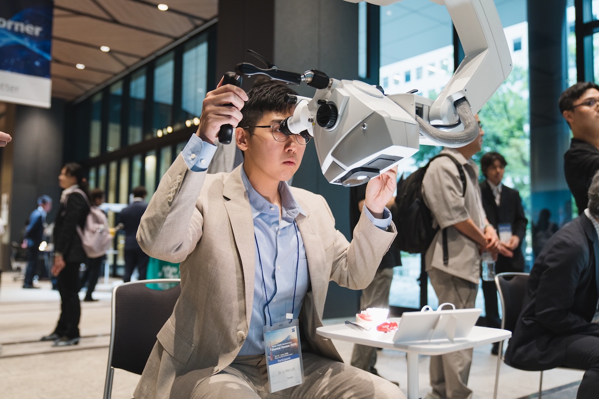
在蒔美牙醫集團,顯微根管治療的應用旨在透過儀器輔助提高臨床檢查與操作的清晰度,主要特點包含:
At Smile Dental Group, microscopic endodontic treatment aims to enhance clarity in clinical examination and procedure execution through instrument assistance. Key features include:
提供充沛照明與放大視野
蔡司顯微鏡具備高倍率放大與同軸光源,能照亮狹窄的根管內部,減少視野盲區,協助醫師檢視微小裂隙與管路開口。
Abundant Illumination and Magnified Vision: ZEISS microscopes feature high magnification and coaxial lighting to illuminate narrow canal interiors, reducing blind spots and helping doctors inspect micro-cracks and canal orifices.
注重細節概念,保留健全齒質
在清楚的視野下,醫師切削齒質的範圍更為明確,有助於減少不必要的結構移除,儘可能維持牙齒原本的強度。
Detail-Oriented Approach to Preserve Healthy Tooth Structure: Clear vision allows precise tooth reduction, helping reduce unnecessary structure removal and preserving the tooth's original strength as much as possible.
協助維持良好的密封品質
根管清創後的封填密合度對於預後相當重要,顯微鏡能輔助醫師檢查充填過程的細節,維護療程穩定度。
Assisting in High-Quality Sealing: Sealing tightness post-debridement is critical for prognosis; microscopes assist doctors in checking filling details to ensure treatment stability.
增進醫病溝通與資訊透明度
顯微設備常配備影像紀錄功能,醫師可將牙齒內部的實際狀況呈現給患者了解,協助患者明確掌握自身的口腔狀況與治療計畫。
Enhancing Doctor-Patient Communication and Transparency: Microscopic equipment often features image-recording capabilities, enabling doctors to present real conditions inside the tooth to patients, helping them clearly understand their oral health status and treatment plan.
為什麼顯微鏡輔助對牙髓處置至關重要?
Why is Microscopic Assistance Crucial for Endodontic Procedures?
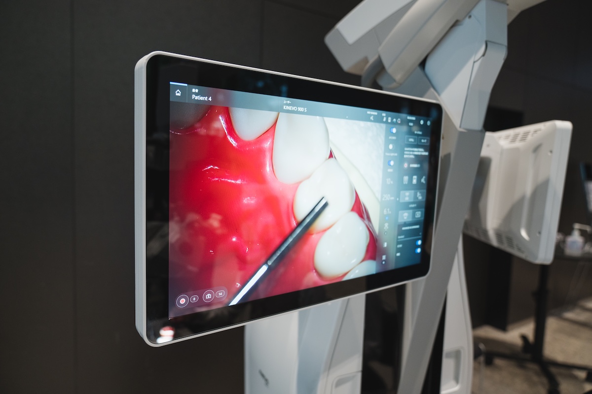
牙齒內部的解剖構造宛如一座複雜的微型迷宮,傳統肉眼觀察往往受限於角度與光線,難以看清深處的細節。過往在沒有顯微鏡輔助的情況下,醫師在面對彎曲根管、鈣化通道或遺漏根管時,許多時候必須依賴手感與經驗進行探索。
The internal anatomical structure of a tooth resembles a complex micro-labyrinth. Traditional naked-eye observation is often constrained by angle and lighting, making deep details difficult to see. In the past, without microscopic assistance, doctors dealing with curved canals, calcified passages, or missed canals frequently had to rely on tactile feel and experience.
顯微鏡的導入,將「盲操作」轉變為「直觀視野」。當視野放大數倍至十數倍後,牙髓腔內的任何微小變化都得以清晰顯現。這不僅有助於減少誤傷健康牙本質的風險,更能提高清潔與處置的徹底程度。對於面臨複雜狀況或反覆發炎的牙齒來說,顯微輔助處置提供了重新評估與保留患牙的臨床選擇。
The introduction of microscopes transforms "blind procedures" into "direct vision." When vision is magnified several to a dozen times, any minute change inside the pulp chamber is clearly revealed. This not only reduces the risk of accidentally damaging healthy dentin, but also enhances the thoroughness of cleansing and treatment. For teeth facing complex conditions or recurrent inflammation, microscopic assistance offers a clinical option to re-evaluate and save the tooth.
蒔美牙醫集團:重視醫療品質,陪伴患者維護口腔健康
Smile Dental Group: Prioritizing Medical Quality, Accompanying Patients in Oral Health Preservation
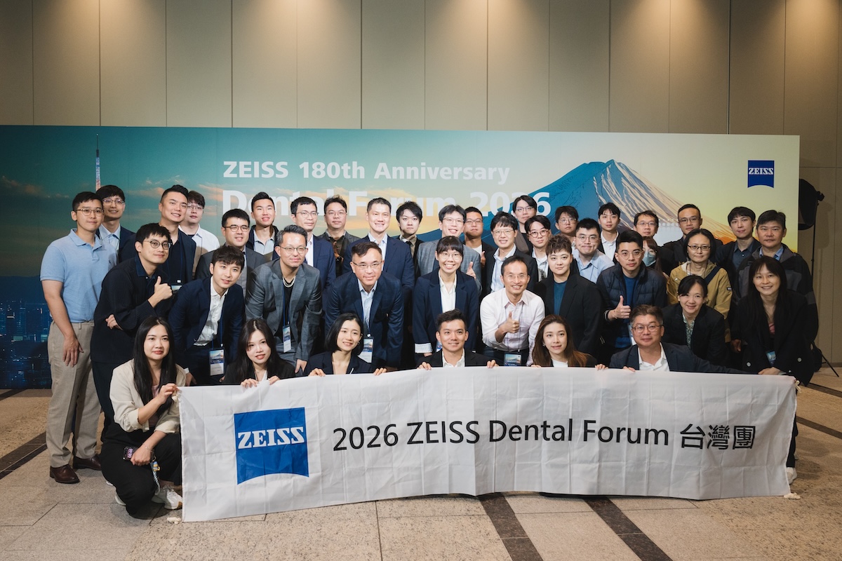
「ZEISS Dental Forum 2026」東京論壇的舉辦,展現了全球牙科醫療持續朝向高放大倍率與細緻化發展的趨勢。蒔美牙醫集團關注國際臨床技術與觀點的推陳出新,並將這些理念落實於日常診療之中。
The "ZEISS Dental Forum 2026" in Tokyo highlights the global dental trend toward high magnification and precision care. Smile Dental Group closely follows innovations in international clinical techniques and concepts, applying these principles to daily practice.
針對複雜的根管狀況或曾面臨治療難題的患牙,顯微鏡根管專科劉立偉醫師與蒔美醫療團隊將以嚴謹的態度,評估客觀條件並提供適當的處置建議。
For complex root canal conditions or teeth that have faced treatment challenges, endodontic specialist Dr. Liu Li Wei and the Smile Dental medical team maintain a rigorous approach to evaluate objective conditions and offer appropriate treatment recommendations.
未來,蒔美牙醫集團也將持續導入適合的醫療設備,落實學術交流與臨床實踐,陪伴每一位患者建立長遠且穩固的口腔健康。
In the future, Smile Dental Group will continue to introduce appropriate medical equipment, foster academic exchange and clinical practice, and support every patient in building long-lasting, stable oral health.